
Cataracts are thought to be synonymous with ageing process. There are different types of cataracts, and most do develop slowly. However, posterior subcapsular cataracts result in faster deterioration of vision in comparison to the other types. However, they are treatable, thanks to advancements in the field of ophthalmic surgery.
Symptoms of Posterior Subcapsular Cataracts
You should be able to observe symptoms within the first few months of their advent.
- Each eye lens is nested within a lens capsule. It is a membrane or small sac. The capsule ensures that the lens stays in position.
- The regions adjacent to the anterior and posterior capsules are referred as subcapsular regions.
- The posterior subcapsular area is towards the back/posterior of the lens. Therefore, clouding/opacification in this part of the lens is called posterior subcapsular cataract.
- Similar to other cataracts, the opacity is due to the clumping of protein fibres on the lenses.
- Since the dense clumping blocks the path of light, you may find difficulty with reading.
- You are not able to tolerate brilliant sunlight either as this type of cataract leads to greater scattering of light.
- Bright lights tend to be too glaring or depict halos around them, specifically at night.
- Your eyes feel comfortable with dim illumination only.

Figure 1. Posterior subcapsular cataract.
Causes of Posterior Subcapsular Cataracts
Who is most at risk for succumbing to this condition?
- Age is the commonest risk factor
- Exposure to radiation
- Diabetes mellitus or other systemic conditions
- Overuse of corticosteroids
- Electric shock
- Sufferers of retinitis pigmentosa, intraocular inflammation/ uveitis
- Blunt trauma to the eye
- Extreme nearsightedness
- Someone who has undergone vitreoretinal surgery
How do posterior subcapsular cataracts develop?
- The entire lens of each eye is a mass of fibres and water.
- The fibres comprise of proteins.
- The intelligent and uniform arrangement of lens fibres is responsible for transparency of the lens that allows light to enter the eye and come to a focus on the retina..
- With age and due to other risk factors described above, these fibres become overactive.
- There is new growth below the lens capsules.
- Since they need their own space, they just scatter the older fibres.
- Thus, a cluster of new fibres begins to form in the subcapsular region. This results in visual disturbances.

Figure 2. Phaco probe used to perform cataract surgery (phacoemulsification)
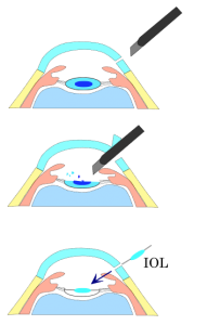
Figure 3. Phacoemulsification procedure.
Treatment of Posterior Subcapsular Cataracts
Thick glasses or good-quality bifocals may work for a while. You may even try the latest visual aids or prefer to use a magnifying lens. However, do not expect the ‘good news’ to last for long. You will have to go for surgery! The medical fraternity refers to it as phacoemulsification.
- Modern cataract surgery or phacoemulsification involves usage of an ultrasound probe.
- The sound emitting from it suffices to break up (emulsify) the cataract.
- The doctor removes all the bits and pieces with the aid of a tubing attached to the phaco machine. This suctioning tube behaves like a vacuum cleaner.
- Once the cataractous material within the capsule becomes clean, the surgeon places a foldable intraocular lens into it.
- The entire surgery takes about 10 minutes and is most commonly done under topical anaesthesia i.e. no injection no stitch cataract surgery. The patient walks in and walks out of the operation theatre.
- This is a day care procedure and patient is prescribed eyedrops for about 3 weeks after the procedure.
- According to the type of intraocular lens implanted in the eye, the patient may or may not require prescription glasses after surgery.
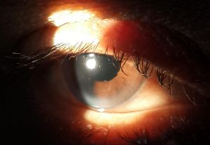
Figure 4. Postoperative picture after implantation of intraocular lens.
- High Blood Pressure and Vision: How Your Eyes Show the Warning Signs - May 27, 2026
- Can the 20-20-20 Rule Protect Your Eyes from Digital Strain? - May 27, 2026
- Can Swimming Cause Eye Infections? Summer Eye Safety Tips - April 27, 2026


