Distorted or blurry vision is alarming, and it may happen if you develop a macular hole. It is a tiny gap that opens up in the macula area located at the center of the retina.
The retina is a light-sensitive layer (neurosensitive tissue) which is the innermost layer of the eye. It converts light signals into electrical signals that are carried to the brain. The center part of the retina is called macula and is responsible for clear and detailed vision. The retina is composed of several layers each having a definite function. The majority of the retina has photosensitive cells known as ‘rods’ while the macula has the highest density of ‘cones’. The macula is responsible for the fine-detailed vision for carrying out daily tasks like reading and color vision, recognizing faces, driving, etc.
What Causes Macular Holes and Whom It Affects?
Generally, macular holes are caused by the age-dependent contraction of the vitreous jelly and can be partial or even full in thickness. Macular holes can vary in size. As the vitreous substance shrinks, it can pull the retina.
In many cases, the vitreous layer separates cleanly from the retina. However, in some people, it is tightly stuck and tears a hole in the macula. Also, the retinal fibers present on the surface can further pull on the hole and enlarge it.
When compared to men, women are at a slightly higher risk of developing macular holes. Generally, macular holes are related to aging processes. People are likely to develop macular holes in the later stages of life when they are 60 or above.
When a macular hole develops, the majority of the people notice a decrease in vision. In the initial stages, macular holes cause blurred and distorted vision. Many patients complain of trouble reading the small print, and straight lines appear to look wavy or bowed. Then, people see a ‘missing patch’ or small black patch in the center of the vision.
Conditions That Can Lead to Macular Holes
Macular holes can occur due to:
- A detached retina
- Diabetic eye disease
- Vitreous shrinkage
- Vitreous separation
- Myopia
- Macular pucker
- Eye injury

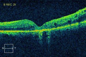
OCT scan of lamellar macular hole
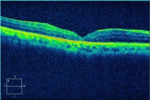
Post-operative OCT scan of the same patient with lamellar macular hole
Treatment Options for Macular Holes
Usually, to treat full-thickness macular holes, surgery is needed. ‘Vitrectomy’ is the surgical procedure to treat macular holes. This surgery is often successful and can help prevent further vision loss. In many cases, vitrectomy has resulted in improved vision.
The macular surgery involves removal of the vitreous jelly (vitrectomy) which helps to stop it from pulling on the retina. After removing the vitreous internal limiting membrane (acellular superficial basement membrane of the retina) surrounding the macular hole is peeled to relieve the pulling forces (traction) causing the macular hole to form and enlarge. At the end of the surgery, the vitreous cavity is filled with special long-acting gas. The surface tension of the gas bubble helps in closing the macular hole. The macular surgery is done under the influence of local anesthesia and following the procedure patient is expected to stay in a face-down position. This positioning is necessary for maximum up to three days. Maintain this positioning so that the gas bubble can press against the macula. In case you have a problem in maintaining the face-down position one could lie in a sidewards position. Face up a position with face facing the ceiling is best avoided for the initial three days after macular hole surgery.
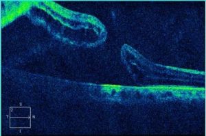
OCT scan of the full-thickness macular hole with retinal detachment
OCT scan of the same patient with full-thickness macular hole and retinal detachment
Precautions to Take After Macular Surgery
The doctors at Neoretina advise patients to avoid air travel for about two weeks. This is because the gas bubble in the eye can expand owing to changes in air pressure. Air travel can increase eye pressure.
After the macular surgery, the chances of cataract or clouding of the eye lens is common. This can call for another surgery in which the clouding of the eye lens is removed, and the lens is replaced with a clear plastic intraocular lens. As per Neoretina specialists, this is not an issue in the patients who have already gone through cataract surgery.
Improvement of vision following macular hole surgery varies from one patient to another. Patients with a macular hole for less than six months have good chances of recovering their vision than those with longer duration. Also, people with a smaller size of macular holes have better results.
- Retinitis Pigmentosa Symptoms, Causes, and Treatment - September 23, 2019
- Diabetic Vitrectomy: A Quick Guide - June 7, 2019
- Macular Surgery: What You Should Know - June 5, 2019