Optical Coherence Tomography (OCT) in ophthalmology is an emerging technology and is used to obtain high-resolution cross-sectional images of the nerve layer on the inside of the eye, the retina. It is a non-invasive imaging technology. This technology provides images on the micron scale in real time. The retinal layers can be differentiated, and the thickness of the retina can be measured with optical coherence tomography.
With the help of such sharper and clearer results, an ophthalmologist can detect and diagnose various retinal diseases and also emerging problems early. With the latest swept-source technology faster images can be obtained and even the structures behind the retina, ie. choroid (vascular layer of the eye) can be visualised.
Very few centres in the country have acquired OCT angiography, which is a no dye, no injection technology used to analyse blood flow in the retina and choroid. Since this technology does not involve injection of a dye, complications related to dye injection like anaphylaxis (severe allergic reaction), vomiting etc are totally obviated. This technology is especially useful in evaluation of patients with kidney disease who are not suitable for fluorescein dye injection.
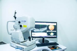
Swept source OCT with OCT angiography machine
Today, optical coherence tomography has become the most comfortable and standard procedure for the assessment, diagnosis, and treatment of several retinal and uveal conditions and glaucoma. This technology uses light rays to measure the thickness of the retinal tissues. In the OCT scan, no X-rays or radiations are used. It is a non-contact scan and therefore it does not hurt nor does it make patients uncomfortable. It can be acquired in less than a minute.
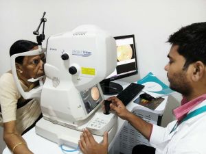
Optical Coherence Tomography during an Eye Examination
Optical Coherence Tomography helps ophthalmologists to view the anatomical structures of the back of the eye. With this technology, an eye professional can take clear and detailed images of the various structures situated at the back of the eye including retina, optic nerve, macula, and choroid. It works by using interferometry which enables an ophthalmologist to image the tissue with the help of a beam of light directed into the eye. Some part of this shining light is reflected by different eye tissues. On the basis of this, images are built.
OCT scan in progress
Uses of Optical Coherence Tomography
Your eye doctor may advise you for an Optical Coherence Tomography scan for a plethora of reasons which include:
-
Diagnosis of various eye conditions like a macular hole, macular oedema, macular pucker, glaucoma, age-related macular degeneration, central serous retinopathy, vitreous traction, diabetic retinopathy, etc.
-
Monitoring of eye diseases like uveitis.
-
Discounting or verifying suspected retinal swelling.
-
To check the effectiveness of the given medication regime.
-
To check or compare OCT results against visual acuity.
-
In recent times, Optic Coherence Tomography is used in graft assessment after corneal surgery.
-
OCT is also used to evaluate the conditions related to the optic nerve as a scan to observe changes to the fibres of the optic nerve.
Indications of Optic Coherence Tomography
The indications of Optic Coherence Tomography have increased during the past few decades. Recently, OCT is widely used in various ophthalmic diseases which affect retina of the eye, the angle of the eye, uvea (vascular middle layer of the eye) and cornea. The non-contact nature and high acquisition speeds of this new technology offer a wide range of quantitative and qualitative information. With Optical Coherence Tomography imaging clear and detailed images of in-vivo examination of human eyes can be obtained.
For Optical Coherence Tomography, the dimension of the cataract along with its shape, the intraocular lens position, the posterior capsular bag, and the incision healing have become the new potential targets. Also, OCT and OCT angiography have given insights into the study of natural history of diseases of the retina and pathophysiologic mechanisms of healing in diseases of the choroid. Thus, OCT is a promising technology which can help ophthalmic research as well as clinical practice.
- Retinitis Pigmentosa Symptoms, Causes, and Treatment - September 23, 2019
- Diabetic Vitrectomy: A Quick Guide - June 7, 2019
- Macular Surgery: What You Should Know - June 5, 2019