One of our most crucial and valuable senses is ‘vision’. Eyes are made up of several parts and all of them are equally important for vision. When light enters into the eye, it is directed in the right amount towards the eye lens. The complex anatomy of the eye converts this incoming light into electrical energy.It is then transferred to the brain through electric impulse where it is processed for vision.
1. Understanding the eyeball– Iris, Ciliary body and layers
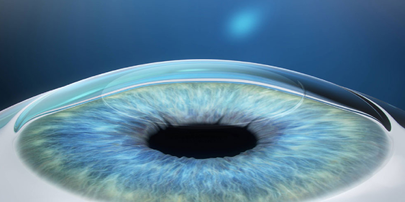
To see things normally, many important eye structures are involved which include (Figure 1):
Cornea: At the front of the eyeball, there is a circular, transparent protective layer called the ‘cornea’. It refracts the light entering into the eye lens. The cornea is sensitive to pain and involuntarily causes closing of the eyelid if anything touches it.
Sclera: It is the tough, protective white part of the eye.
Pupil: Light enters into our eye lens through this small circular opening located at the center of the Iris. Constriction or dilation (narrowing or widening) of the pupil is controlled by the Iris.
Uvea: It is the middle, pigmented layer of the eyeball. Uvea consists of three segments- the iris, the choroid, and ciliary body.
- Iris: This structure acts like a diaphragm of a camera. Iris is the visible, coloured part of the eye and is located right in front of the lens.
- Choroid: the middle layer of the eye, between the retina and sclera, it consists of connective tissues and blood vessels. It is situated between the retina and the sclera. This important structure provides nutrients and oxygen to the retina.
- Ciliary body: This structure connects the iris and the choroid and plays a major role in accommodation which is required for clear vision at all distances. It also secretes aqueous humour, which maintains the eye pressure and provides nourishment to eye.
- Lens: Enclosed in a thin transparent capsule, the lens is a transparent structure. It is located behind the pupil. To focus on objects situated at different distances or locations, eye lens can change its shape. With age, the lens becomes thicker or cloudy which can make focusing on objects a bit difficult.
- Retina: Retina is the light-sensitive layer of the eye. It has millions of light-sensitive cells known as ‘photoreceptors’ or ‘rods and cones’. Rods functions when the light is dim while the cones in the bright light. Cones are of three types and each is sensitive to the wavelength of a distinct primary colour- green, red or blue.
- Macula: It is the central, most sensitive part of the retina, located at the back of the eye. It surrounds the fovea.
- Fovea: It consists of a huge concentration of cone cells and is responsible for accurate reading vision.

Figure 1. Parts of the human eye
2. Role and functioning
Eye is one of the most complex organs of the body and it is responsible for carrying out crucial functions. The basic role and functioning of various eye structures are dedicated to offer clear vision. Iris is an important structure which regulates the quantity of light that enters the eye. The Choroid contains a pigment that absorbs excess light and prevents blurring of vision. Lens refracts light and focuses it on to the retina. The light sensitive retina has photoreceptors that capture the light that falls on it. These photoreceptors convert the signals into electrical impulses. Through the optic nerve, these signals are then sent to the brain’s visual areas.
Apart from focusing on objects, perceiving colour and detail, eyes are responsible for producing tears which nourish and lubricate the eye surface and wash away debris.
3. Mechanism of eye inflammation
Inflammation is the natural defense response of the body to any damaged tissue, toxin, foreign body or germs. But inflammation also brings with it some tissue damage. Eye is unique as it is an immunologically privileged site. This immune privilege makes sure that eye is protected from day to day inflammatory insults. Therefore, the integrity of the various eye structures is maintained and they are able to provide clear vision. When there is a breach in this protective micro-environment of the eye, Uveitis (eye inflammation) is said to occur. This leads to breakdown of the blood-eye-barrier and structures of the eye can now be damaged by inflammation, thus causing loss of clear vision. Eye inflammation can occur due to numerous causes such as bacterial or viral infections, autoimmune rheumatic diseases, etc. The prevalence of Uveitis is reported to be 204/100,000 population. It is a major cause of irreversible blindness. Every year 17.6% patients with uveitis develop temporary or permanent vision loss. About 12.5% develop glaucoma.
4. Secondary conditions
Uveitis is an inflammation of the uveal tract or uvea (middle pigmented layer of the eye) but it can also damage adjacent structures like the retina, vitreous and sclera.
If eye inflammation needs to be treated in time and it can cause secondary eye conditions like (Figure 2):
- Damage to the cornea (front transparent part of the eye)
- Cataract (clouding of the lens of the eye)
- Glaucoma (optic nerve damage due to eye pressure)
- Scarring in the retina
- Retinal detachment.
- Bleeding in the eye
- (Macular edema) Swelling of the centre of the retina
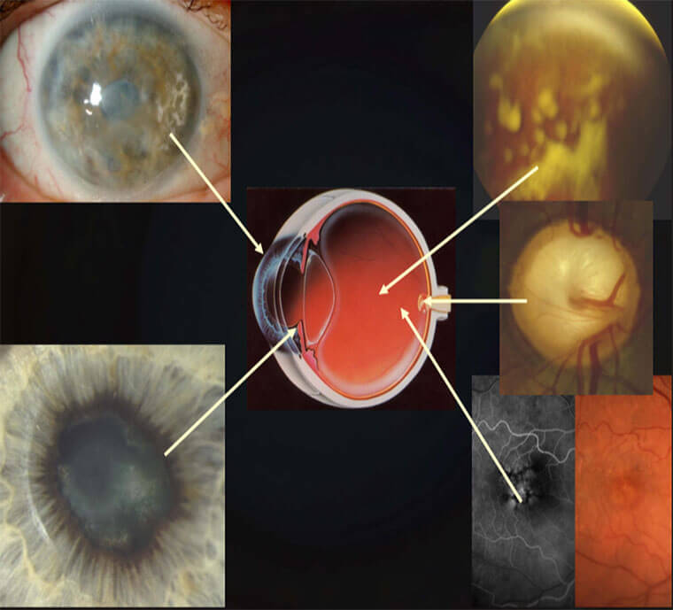
Figure 2. Complications of uveitis
5. Watch for signs
Although the signs and symptoms of eye inflammation vary according to the major site of inflammation in the eye, some of the characteristic symptoms include (Figure 3):
- Redness
- Decreased or changed vision
- Eye pain
- Floaters
- Light sensitivity
In case of Uveitis, symptoms may occur suddenly and can get worse in no-time. In some people, symptoms develop gradually and may affect either one or both the eyes.

Figure 3. Symptoms of uveitis
Treatment of Uveitis
Infectious uveitis is easily treated with the appropriate dose of anti-microbial tablets and steroid eye drops with or without steroid tablets. For the treatment of non-infectious Uveitis stepladder approach is used as it tends to occur recurrently. Generally, to treat the auto-immune or non-infectious uveitis, steroid drops are prescribed first. Then, the treatment is gradually advanced to steroid injections and/or tablets. The steroid medication helps reduce eye-inflammation.
If the uveitis continues to reoccur when a steroid medication is stopped or reduced in dose/frequency, systemic immunosuppression with an immunomodulatory chemotherapeutic drug is instituted.
Medication should only be taken as per the doctor’s advice. Patients suffering from uveitis should never start or stop taking the medication on his/her own. The treatment of uveitis is generally prolonged and it needs acceptance, persistence, and cooperation from both the patients and their family members. The sooner uveitis is diagnosed, the chances of successfully treating the condition increases. It is always advisable to consult a specialist in a reputed eye care hospital to take the right call on the course of the treatment for this serious eye disease.
- Choosing the Right Intraocular Lens: Make an Informed Decision - February 28, 2019
- Intravitreal Injections: Indications, Procedure, Do’s, Don’ts and Myths - February 13, 2019
- Cataract Surgery, Post-Operative Care | Neoretina - February 13, 2019