The retina is the thin layer of tissue that is light-sensitive. This layer lines the inside of the back of the eye. It sends visual messages to the brain via the optic nerve. In some people, the retina detaches; it is pulled or lifted from its normal position. Retinal breaks or tears in small retinal areas can result in retinal detachment.
About Retinal Detachment
As retina is needed to see properly, it is crucial to treat retinal detachment without delay. Typically, it is a progressive condition in which the retina has one or more holes/ tears or it detaches from the retinal pigment epithelium. This allows the subretinal fluid to pass and accumulate underneath them. This causes the thin layer of the retina to detach from the tissues present in the back of the eye. When this retinal layer becomes loose or separated, the vision can become blurred or distorted.
In some patients, small blood vessels also bleed into the vitreous and further cloud the vision. This eye condition needs prompt treatment; otherwise, it causes permanent vision loss. Mostly, retinal detachment affects one eye; however, in some patients both the eyes can get affected.
Types of Retinal Detachment
There are three types of retinal detachment
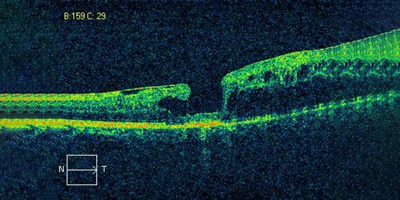
Rhegmatogenous: (Figure 1) It is the most common type and involves a retinal break or tear. This allows fluid to seep under the retina. This leads to the detachment of the retina from the back wall of the eye, also known as ‘retinal pigment epithelium’ that nourishes the retina. Retinal tears are formed due to an event called posterior vitreous detachment. Vitreous gel is adhered to the retina on its entire surface area. At some areas its is more firmly adhered to the retina especially areas of thinning called lattice degeneration than in the rest of the retina. With age the vitreous gel liquifies and causes the separation of the vitreous from the retina called posterior vitreous detachment(PVD ). During the process of PVD i.e ,separation of vitreous from retina there are chances of retinal tears formation at the areas of tight adhesion of vitreous and retina. These retinal tears if not treated can go on to cause retinal detachment

Figure 1. Rhegmatogenous retinal detachment.
Tractional: (Figure 2) In this type, the scar tissue which is present on the surface of retina contracts and pulls it away from the retinal pigment epithelium. This is more frequently seen in advanced stages of diabetic retinopathy, and is called proliferative diabetic retinopathy where new vessels develop in the eye which leak protein forming membranes which in turn pull the retina causing retinal detachment.

Figure 2. Tractional retinal detachment.
Exudative: A rarer type is the exudative retinal detachment. It is caused by retinal disease like inflammatory disorders,. In the exudative type, the blood vessels underneath the retina leak fluid but cause no retinal breaks or tears.
Signs and Symptoms
The first common signs and symptoms of retinal detachment include loss of field of vision, the sudden appearance of flashes of light, cobwebs and spots or floaters in the vision, heaviness in the eye, etc. These signs may be followed by a dark ‘curtain’ or dense shadow moving across the vision. Eyesight will be greatly affected if the central macula area of the retina gets detached.
Diagnosis
If you experience any of these signs or symptoms, book an appointment and seek advice from an expert within 24 hours. Getting medical help as soon as possible lessens the chance of permanent eyesight damage. For the diagnosis, your ophthalmologist will first examine your eyesight. Depending on your condition, your ophthalmologist will carry out the following investigations:
- visual acuity testing
- slit-lamp examination
- indirect ophthalmoscopy
- B-scan ultrasonography of the affected eye
Risk factors
Retinal detachment is very common among the middle-aged and the elderly. Some other risk factors include:
- Blunt trauma
- People with high myopia
- People who have had cataract surgery
- Risk of developing detachment is more in those individuals who are or were affected with a retinal detachment in the other eye.
Rarely, some types of retinal detachments run in families.
Treatment and Aftercare
There are several methods like Cryopexy and Laser Photocoagulation, Scleral buckle surgery, Pneumatic retinopexy and Vitrectomy for treating a detached retina. (Figure 3) After-care is important for the speedy recovery after retinal detachment surgery. Your ophthalmologist will direct you about aftercare needs in detail. Follow the instructions of your surgeon or ophthalmologist which typically include:
- Taking medication
- Taking rest
- Avoiding the rubbing of eyes, etc.

Figure 3. Retinal surgery in progress at Neoretina.
Prevention
Early diagnosis of retinal detachment is the key to prevent blindness or vision loss. Hence, it is crucial to report immediately to a retina specialist in case of any symptoms described above
If you notice any sign and symptom of retinal detachment, contact a specialist or a NABH accredited eye care hospital today.
- Retinitis Pigmentosa Symptoms, Causes, and Treatment - September 23, 2019
- Diabetic Vitrectomy: A Quick Guide - June 7, 2019
- Macular Surgery: What You Should Know - June 5, 2019