Envision Diagnostics
State-of-the-Art Diagnostic Facility
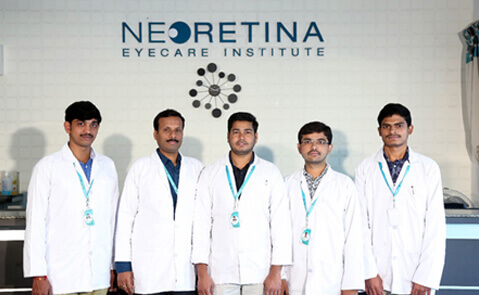
Team
Team of highly skilled optometry staff for performing and reporting scans,
led by Mr. Nagesh Vuppala, M. Optom
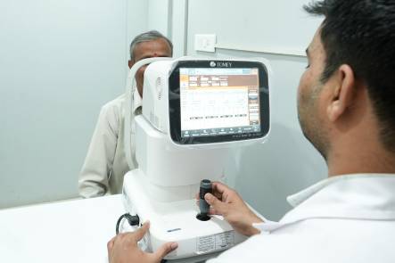
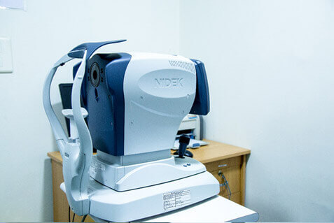
Tomey OA 2000 optical biometer:
In order to provide precision cataract surgery to our patients, Neoretina has now acquired the Tomey OA-2000 optical biometer. It is a complete system for modern intraocular lens power calculation. It combines high speed biometry measurement with a deep penetration for very dense cataracts and topography. It has the following excellent features:
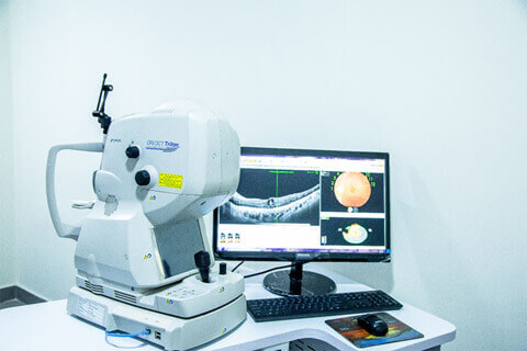
We are proud to introduce for the first time swept source OCT angio machine outside institutional setup in the city of Hyderabad and the states of Telangana and AP. It detects motion contrast and identifies blood flow in vessels by detecting difference in signals in multiple successive scans.
Scan rates have increased from 7000 A scans per second in time domain OCT to about 1,00,000 A scans per second in the current generation of Swept source OCT thereby providing higher resolution of OCT.
Advantage of SSOCTA over FFA:
Advantage of SS OCTA over Spectral domain OCT (SD OCT):
SS OCT works at a wavelength of 1050 nm while SD OCT works at a wavelength of 840 nm. Due to higher wavelength, SS OCT can
It is comprehensive too.
SSOCTA report provides comprehensive picture showing angiography report of different layers (Superficial capillary network, deep capillary network, outer retina and capillary network of choroid), a high quality SS OCT image and a fundus image. This helps in comparing the various aspects of the OCT simultaneously thereby making better interpretations
This machine can also be used to image anterior segment as well as in glaucoma for measurements of angles and disc angiogram and nerve fibre layer analysis
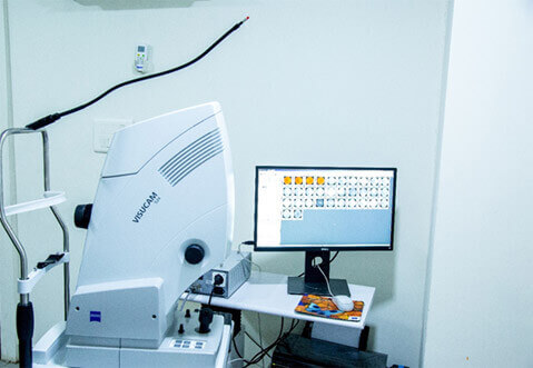
We are equipped with the latest generation Carl Zeiss Visucam 524 machine which has a simple design and is easy to handle and provides ultra high resolution(24 megapixels) fundus imaging in multiple modalities including FFA, ICG and fundus autofluorescence.
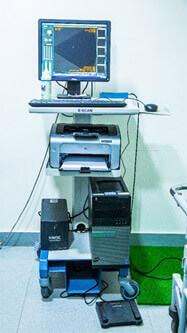
We are equipped with both Ultrasound B scan and UBM. B scan helps in imaging the posterior segment of the eye while UBM helps to image the pars plana, ciliary body and the anterior chamber of the eye. This helps in identifying both posterior segment disorders as well as anterior segment pathologies.
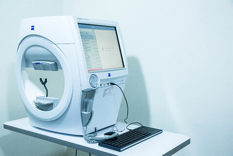
The new Zeiss HFA3 comes with the latest Liquid Lens Technology which enables faster examination by automatically loading the patient’s refractive correction from their previous exam. This also reduces chance of error when selecting a lens during test setup.
It has an improved gaze tracking initialization and easy to use kinetic graphical user interface with full 180° testing range.
It measures progression simply and easily with Guided Progression Analysis (GPA). RelEYE helps to preserve an image of the patient’s eye at every stimulus presentation during a SITA Standard test.
It checks for ptosis or trial lens rim artifacts as well as assess patient's compliance with RelEYE in both GPA and single-field analysis modes.
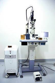
Envision has one of the most advanced LASER delivery system for posterior segment disorders. The LASER delivery system has multiple wavelengths thereby catering to varying indications. While green LASER is traditionally the first choice of treatment, yellow LASER has some inherent advantages like
Similarly, the advantages of multisport LASER over the conventional single spot LASER include
The entire 3 sittings of PRP can be concluded in a single session if required in certain deserving situations

We are equipped with the best YAG delivery system and is suitable for indications like YAG capsulotomy and YAG iridotomy. It ensures optimum amount of energy being delivered to the target tissue resulting in minimal amount of energy being needed for photodisruption and high precision treatment.
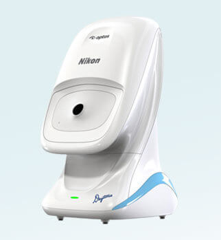
With the acquisition of the Optos ultra-wide field retinal photography camera, Neoretina Eyecare Institute becomes the first super speciality private hospital in Telangana to offer this technology to patients. The images captured by Optos will enable us to monitor and document the patient’s eye condition from a visit to visit. It’s cutting edge technology offers the following advantages:
Practice Efficiency: Optos enables faster imaging, gives a detailed 200º photograph of the retina, as against the previous view of only 50º, thus improving the diagnosis. Images are acquired through a 2mm pupil (without the need for dilatation) and even through media opacities like cataract. This helps the clinician make a clear diagnosis in a wide range of ocular diseases.
Improved Clinical Decision-making: Unlike conventional equipment, Optos signals early signs of many ocular diseases in the retinal periphery. This speeds up treatment planning and chances of recovery.
Improved Chances of Vision Recovery: Enables the specialist to detect eye abnormalities sooner and manage them effectively.
“I was highly impressed by the over all management of large number of patients properly guided and investigated very effectively. Staff at the reception and all management levels well trained and helpfully caring hardworking. My salute to all involved for starting proper management of large number of patients.”
SAROJINI UGALE
“The staff was very much kind and the clinic setup was very beautiful. Very nicely managed. I definitely would like to recommmed this clinic to my near and dear ones. Thanks once again for taking care of me!”
AZAM KHAN
“Excellent in all respects. Very clean and has well maintained toilets. The staff are very friendly and helpful. Overall extremely satisfying experience.”
SASIDHARA REDDY
“My dad had a retinal detachment which was found overnight and he had to undergo retinal detachment surgery (vitrectomy) within 7days. He flew down to Hyderabad and directly to Raja Ram Reddy. He suggested to go for surgery the very next day. It was a successful surgery and my dad got discharged in the evening. He is very cordial and really nice in speaking. He is calm cool minded. A perfect doctor to consult.”
NEHA PATNAIK
“I'm very happy with the drs approach and very patiently examined I have great confidence in Dr reddy its been three years that I come to Dr reddy all the way from Kolkata wish we have doctors like him at Kolkata too.”
ANIL KUMAR AGARWAL
“Best..Wonderful clinic with very helpful staff and well experienced and caring doctors like Dr.Srinivas and Dr.R.R.Reddy... I took my father who is diabetic and heart patient also for his eye surgery and the doctors guided us v.nicely to take care of him b4 & after surgery and my father's eyes r perfect...mashallah...thanks to God and then the neoretina team...Well done...keep spreading the light of vision in the life of others too. ”
MEHER ZAIDI
“I know her from since 3 years she diagnosed the retina detachment in my left eye and explained about the treatment. She works for NEORETINA eye care institute which is located in the nampally hyderabad. She clarifies each and every doubt of the patient patiently without any irritation. I mean some doctors gets irritated when the patient asks more questions but that's not the case with alka Rani madam. I suggest to all who are suffering from eye disorders to visit neoretina for timely treatment and speedy recovery.”
VASANTHI
“Excellent attention to my eye problem doctor Abhilasha Baharani at Neo Retina Eye Care Institute was very cooperative she is very echo friendly clear voice n determined i will in sha Allah refer to all of my near n dear to visit her if any one having eye problems i appreciate thanks a lot.”
MD MUZAFER
“Dr. Raja is one of the foremost experts in retina issues. He is very approachable at the same time gives you valuable advice.”
T PRASAD